
CORNEAL ULCERS AND NEOVASCULARIZATION
- Sep 14, 2025
- 3 min read
Tavishi
The other day at work, we had a patient with a corneal ulcer. He was here for a recheck, actually, so I didn't get to see the neovascularization, but I did get to hear about it.
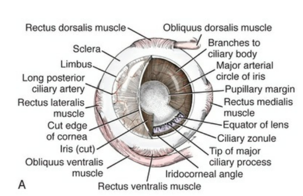
This is a diagram of the dog eye sourced from perhaps the best textbook of all time (Miller and Evan's Anatomy of the Dog). The cornea is the outermost layer of the eye, trapping the liquid and eyebally-parts inside the eyeball, but also, refracting light into the eye.
The cornea consists of several layers: an outer epithelium, Bowman's layer, a stroma (substantia propria), Descemet's membrane, and an endothelium. The outer epithelium is the part that actually faces the outside, and is a squishy living tissue (in science words, non-keratinized stratified squamous epithelium). The tissue allows for gas exchange and prevents stuff from getting in the eye.

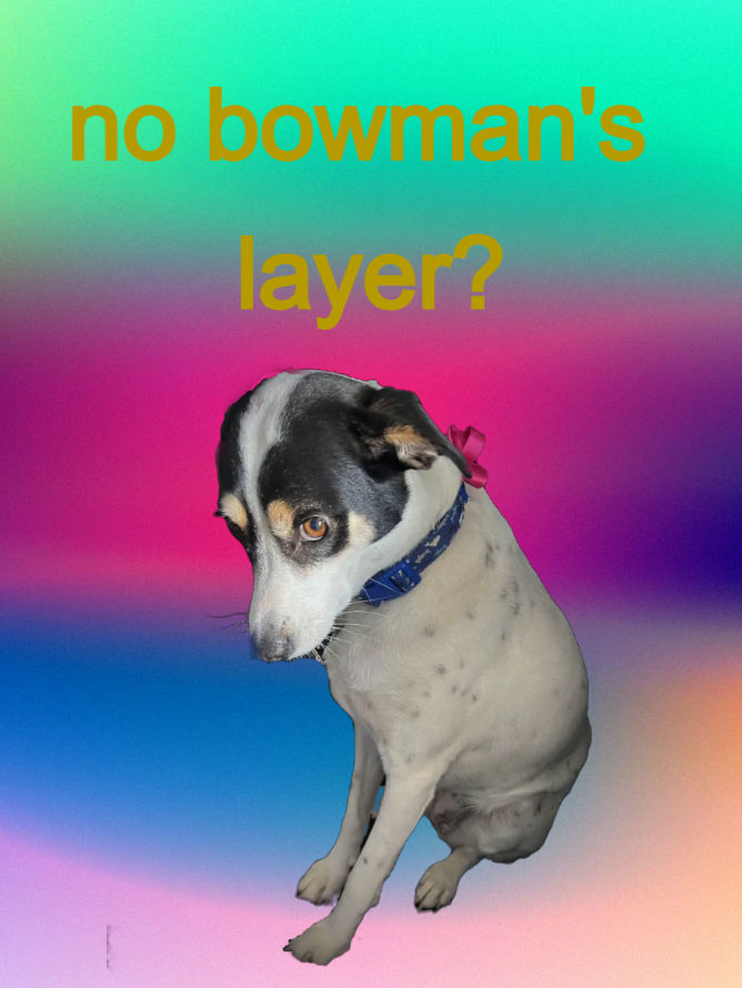
Next, Bowman's layer, which is just kinda like a sheet of collagen and other fibrous proteins between the epithelium and the stroma. Note, the Bowman's layer is not very present in doggies and non-primates. So this dog did Not. have this layer of the cornea.
Anyways, then the substantia propria, which is just collagen and keratocyte soup, and the bulk of the actual corneal tissue.
Descemet's membrane is the final basement membrane separating the substantia propria from real cells, or the corneal endothelium. The corneal endothelium is full of mitochondria-rich squamous cells.

A corneal ulcer is a partial or complete loss of the outer layers of the cornea, and in fancy words, is called ulcerative keratitis. (Sidebar: any time I hear the word ulcerative, my brain autofills colitis.)

Ulcerative keratitis can be classified based on the depth of inflammation. Superficial ulcers penetrate only the corneal epithelium, whereas deep ulcers go as far as Descemet's layer, in which case, they're called descemetoceles.

Corneal ulcers are most commonly caused by blunt trauma, and are most prevalent in brachycephalic dogs like pugs, bulldogs, etc. Those breeds of dogs are also prone to cherry eye (see: horrific genetics). Most of the brachycephalic patients I've seen in my shelter's trauma hospital have eye issues, especially with loss of eye. I think I've only seen one one eyed dog that's not brachycephalic.
Back to the topic at hand: corneal ulcers are identified by three things: cloudiness, neovascularization, and fluorescein stain.
Cloudiness is pretty easily spoken for, but is also a sign of billions of other eye issues, so it isn't always super helpful. Fluorescein stain is really the biggest tell. Drops of an orange fluorescein stain are put into the eye, and then, a blue light is shined into the eye. Whatever areas appear green under that light are ulcers.
Fluorescein is a polar molecule, and doesn't get along well with the cell membranes in the epithelium. However, the stroma is just Cornea Soup, and in areas where the stroma is exposed, fluorescein is able to show up there and look green.

In deeper ulcers, like descemetoceles, because Descemet's layer is hydrophobic, Descemet's layer bulges forward surrounded by a ring of green.
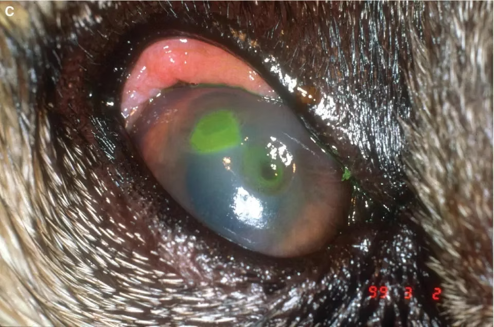
The patient I mentioned at the start of this article displayed neovascularization, which is caused by really cool biochemical pathways. Essentially, the eye freaks out because the eye's super swollen and deprived of oxygen, which means the eye may no longer be clear. And the body's general response to any danger is get blood there right now (see: inflammatory response); so naturally, we get blood to the eye by making new blood vessels.

Neovascularization happens when the body freaks out like that, and is the creation of new blood vessels. When the corneal epithelium gets all hypoxic like that, it releases VEGF, or vascular endothelial growth factor. These bind to VEGFRs, which are tyrosine kinase receptors. They're homodimers that dimerize when their ligand binds to them. yada yada yada
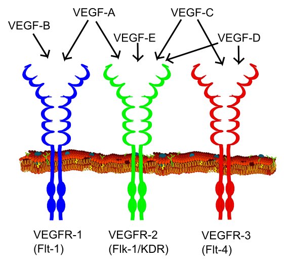
Anyways, the release of VEGF-A is also further induced by the release of HIF, or hypoxia inducible factor. Together, the two factors cause the growth of blood vessels into the cornea. VEGF-A binds to VEGFR-1 receptors on corneal epithelial cells.
All in all, causing the eye to look something like this:

anyways silly little post hope y'all like it :o


Comments